Upper Back Anatomy - Upper Back Pain Anatomy Of The Back The Pain Center Pain Management Care : The extrinsic (superficial) back muscles, which lie most superficially on the back.. The spine is made up of 33 individual bones called ve. The traps) the latissimus dorsi (a.k.a. All these muscles are therefore associated with movements of the upper limb. 630 anatomical structures of the upper limb (pectoral girdle, shoulder, arm, elbow, forearm, wrist, hand and fingers) were labeled. The main superficial muscles of the back are the following:
The spine is made up of 33 individual bones called ve. These muscles facilitate movement by attaching to one or. It is very stiff, and the thoracic spine has a limited range of motion. The rhomboid muscle is activated as you bring and squeeze your scapula or shoulder blades back and together. Related posts of upper back muscle diagram muscle anatomy diagram.
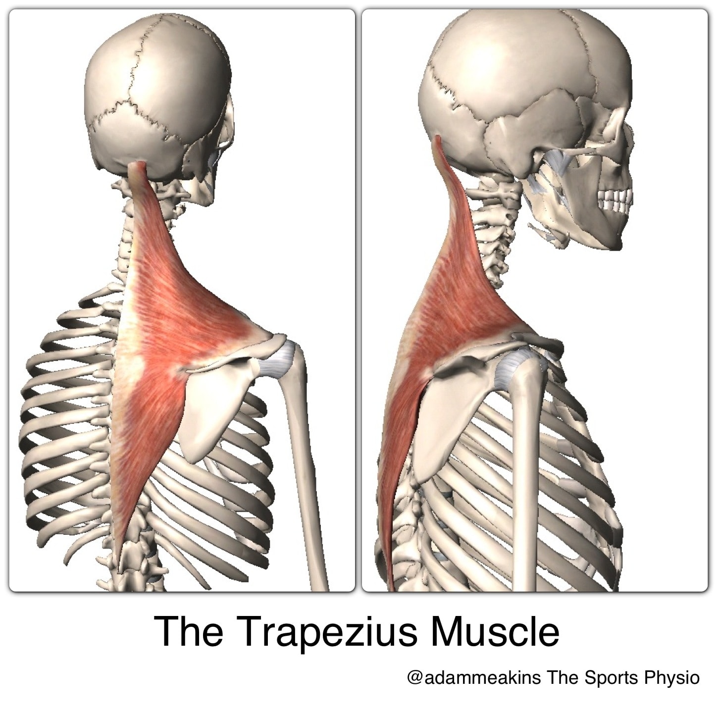
The Upper Traps Over Assessed Over Blamed And Very Misunderstood from www.physiotutors.com Both the deltoid and the trapezius are firmly attached to the spine of the scapula. 1 your spine in this region has a natural inward curve. Balance the weight of your head on top of your spine. The lumbar and sacrum region make up the bone of the lower back anatomy. The extrinsic (superficial) back muscles, which lie most superficially on the back. It runs from the neck to the upper back. This muscle is located on the upper portion of the back anatomy, underneath the trapezius. It is very stiff, and the thoracic spine has a limited range of motion.
Muscle_anatomy_upper_back 4/8 muscle anatomy upper back everything you need to know about the anatomy of the limbs and back and how to apply the material to everyday activities and movements with this updated edition of the classic text.
The trapezius and latissimus dorsi muscles connect the upper limb to the vertebral column. The complexity of this region means that dysfunction can occur either due to injury or progressive pain and degeneration. The human spine is composed of 4 sections of vertebrae. The bones have a crystalline it, essentially, floats off of the back of the chest, as it is connected to the body primarily by muscle. The spine is made up of 33 individual bones called ve. Human anatomy · july 23, 2016. The cervical spine protects the nerves connecting to. The deltoid, teres major, teres minor, infraspinatus, supraspinatus (not shown) and subscapularis muscles (not shown) all extend from the scapula to the humerus and act on the shoulder joint. The cause may be poor posture (such as forward head posture) or any type of irritation of the large back and shoulder muscles, including muscle strain or spasms. Try the injurymap exercise app now. The basic anatomy of your upper back by lindsey mcfadden as you're doing your regular upper back stretching exercises , you're probably wondering about the components of your upper back and why it happens to be the most stable part of your spine. This is my video about the muscles of the back. The traps) the latissimus dorsi (a.k.a.
It is like that for several reasons, all of which you can understand by looking at the anatomy of the thoracic spine. Covering an expanse from the neck to the tailbone, the back muscles are responsible for a broad range of functions, from extending the spine to shrugging the shoulders. The lumbar and sacrum region make up the bone of the lower back anatomy. Balance the weight of your head on top of your spine. The cause may be poor posture (such as forward head posture) or any type of irritation of the large back and shoulder muscles, including muscle strain or spasms.
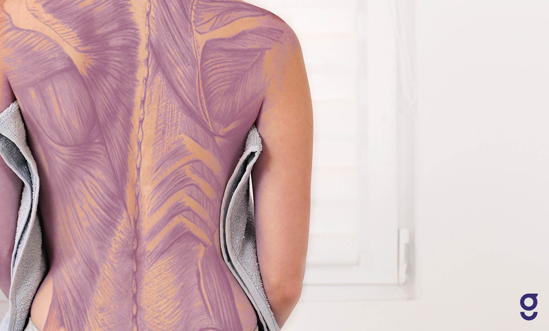
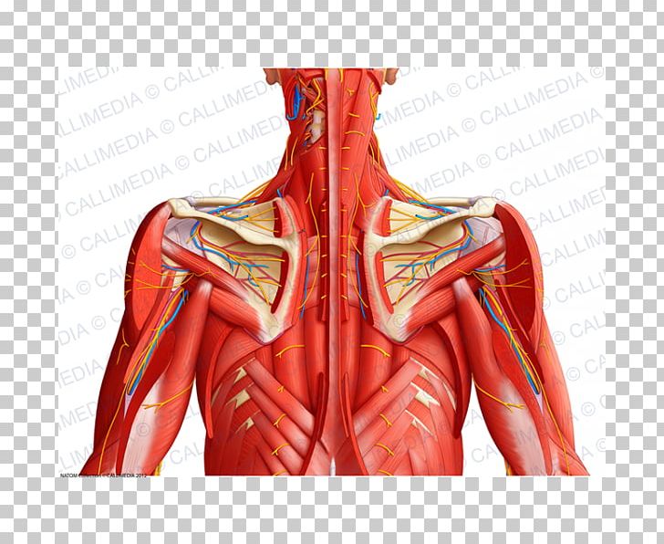
Back Muscles Anatomy Of Upper Middle Lower Back Pain In Diagrams Goodpath from images.ctfassets.net Balance the weight of your head on top of your spine. These sections are cervical (neck), thoracic (upper and middle back), lumbar (lower back), and sacrum (tailbone). In the upper back region, the trapezius, rhomboid major, and levator scapulae muscles anchor the scapula and clavicle to the spines of several vertebrae and the occipital bone of the skull. The traps) the latissimus dorsi (a.k.a. The back functions are many, such as to house and protect the spinal cord, hold the body and head upright, and adjust the movements of the upper and lower limbs. The superficial back muscles are situated underneath the skin and superficial fascia. The lumbar and sacrum region make up the bone of the lower back anatomy. The deltoid, teres major, teres minor, infraspinatus, supraspinatus (not shown) and subscapularis muscles (not shown) all extend from the scapula to the humerus and act on the shoulder joint.
They originate from the vertebrae and insert into the scapulae.
Human anatomy · july 23, 2016. Balance the weight of your head on top of your spine. The bones of the chest and upper back combine to form the strong, protective rib cage around the vital thoracic organs such as the heart and lungs. Powerful muscles that move the head and arms attach to these bones as well. Try the injurymap exercise app now. Upper back pain is most commonly caused by muscle irritation or tension, also called myofascial pain. The cervical spine is the top part of the spine. Back muscles anatomy here include the trapezius, latissimus dorsi, rhomboid and levator scapulae. In the upper back region, the trapezius, rhomboid major, and levator scapulae muscles anchor the scapula and clavicle to the spines of several vertebrae and the occipital bone of the skull. The rib cage also anchors the bones of the head, neck, shoulders, and arms to the trunk of the body. Human musculature bodybuilding infographic muscular system vector human anatomy back muscle anatomy bicep male muscular anatomy human body anatomy female female anatomy muscle hamstrings muscle. The cervical spine supports the weight and movement of your head and protects the nerves exiting your brain. This curve, called lordosis, helps to:
The superficial back muscles are situated underneath the skin and superficial fascia. The cervical spine is the top part of the spine. Balance the weight of your head on top of your spine. Looking for a solution to your back pain problem? 1 your spine in this region has a natural inward curve.
Anatomy Of Upper Back Anatomy Drawing Diagram from cdn.imgbin.com Back muscles anatomy here include the trapezius, latissimus dorsi, rhomboid and levator scapulae. It consists of seven vertebrae. Human musculature bodybuilding infographic muscular system vector human anatomy back muscle anatomy bicep male muscular anatomy human body anatomy female female anatomy muscle hamstrings muscle. The cervical spine protects the nerves connecting to. Evenly distribute weights from your upper body into the lower extremities. The back functions are many, such as to house and protect the spinal cord, hold the body and head upright, and adjust the movements of the upper and lower limbs. This curve, called lordosis, helps to: The bones have a crystalline it, essentially, floats off of the back of the chest, as it is connected to the body primarily by muscle.
Your lower back (lumbar spine) is the anatomic region between your lowest rib and the upper part of the buttock.
The spine is made up of 33 individual bones called ve. The main superficial muscles of the back are the following: Human anatomy · july 23, 2016. Anatomy of the upper back. It is very stiff, and the thoracic spine has a limited range of motion. Both the deltoid and the trapezius are firmly attached to the spine of the scapula. It comprises the vertebral column (spine) and two compartments of back muscles; These muscles facilitate movement by attaching to one or. The trapezius and latissimus dorsi muscles connect the upper limb to the vertebral column. The traps) the latissimus dorsi (a.k.a. The rhomboid muscle is activated as you bring and squeeze your scapula or shoulder blades back and together. The bones have a crystalline it, essentially, floats off of the back of the chest, as it is connected to the body primarily by muscle. The human spine is composed of 4 sections of vertebrae.